
1st PUC Biology Question and Answer: Structural Organisation in Animals
Looking for 1st PUC Biology textbook answers? You can download Chapter 7: Structural Organisation in Animals Questions and Answers PDF, Notes, and Summary here. 1st PUC Biology solutions follow the Karnataka State Board Syllabus, making it easier for students to revise and score higher in exams.
Karnataka 1st PUC Biology Textbook Answers—Reflections Chapter 7
Structural Organisation in Animals Questions and Answers, Notes, and Summary
1st PUC Biology Chapter 7
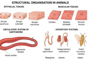
Structural Organisation
in Animals
Scroll Down to Download Structural Organisation in Animals PDF
Question and Answer:
Question 1.
Answer in one word or one line.
(i) Give the common name of Periplanata americana.
Answer:
American cockroach
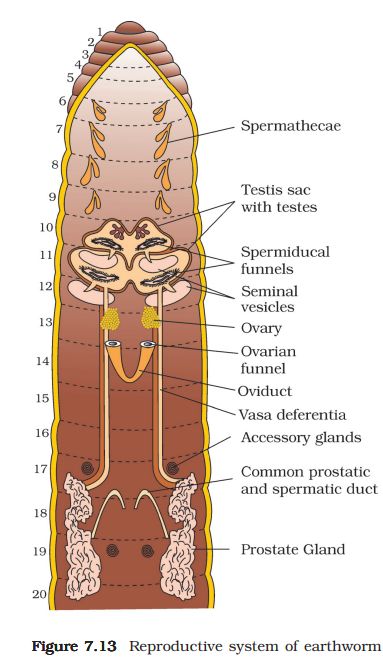
(ii) How many spermathecae are found in earthworm?
Answer:
Four pairs
(iii) What is the position of ovaries in cockroach?
Answer:
Between 2nd and 6th abdominal segments
(iv) How many segments are present in the abdomen of cockroach?
Answer:
Ten segments
(v) Where do you find Malpighian tubules?
Answer:
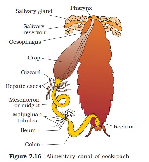
At the junction of midgut and hindgut in cockroach
Question 2.
Answer the following:
(i) What is the function of nephridia?
Answer:
Nephridia are minute tubular excretory organs present in all segments of the earthworm except the first two. Their primary function is to discharge nitrogenous waste and maintain osmoregulation.
(ii) How many types of nephridia are found in earthworm based on their location?
There are three types of nephridia in earthworms:
- Septal nephridia – present on both sides of the intersegmental septa from the 15th segment to the last; open into the intestine.
- Integumentary nephridia – attached to the lining of the body wall from the 3rd segment to the last; open on the body surface.
- Pharyngeal nephridia – present as three paired tufts in the 4th, 5th, and 6th segments.
Question 3.
Draw a labelled diagram of the reproductive organs of an earthworm.
Question 4.
Draw a labelled diagram of alimentary canal of a cockroach.
Answer:
Question 5.
Distinguish between the followings
(a) Prostomium and peristomium
(b) Septal nephridium and pharyngeal nephridium
Answer:
1. Difference between Prostomium and Peristomium
- Prostomium:
The prostomium is a lobe-like structure in front of the mouth. It is sensory in function and helps in burrowing. - Peristomium:
The peristomium is the first body segment (buccal segment) which surrounds the mouth and bears it.
2. Difference between Septal nephridium and Pharyngeal nephridium
- Septal nephridium:
Present on both sides of the intersegmental septa from the 15th segment to the last; they open into the intestine. - Pharyngeal nephridium:
Present as three paired tufts in the 4th, 5th, and 6th segments; they open into the buccal cavity and pharynx.
Question 6.
What are the cellular components of blood?
Answer:
The cellular components of blood are:
- Red Blood Cells (RBCs / Erythrocytes)
- White Blood Cells (WBCs / Leucocytes)
- Platelets (Thrombocytes)
Question 7.
What are the following and where do you find them in animal body.
(a) Chondriocytes.
(b) Axons
Answer:
- Chondrocytes:
These are the cells found in cartilage connective tissue. They produce and maintain the cartilaginous matrix, and their number determines the flexibility of cartilage. They are present in the tip of the nose, pinna of the ear, joints between the vertebrae, etc. - Axon:
It is the longest process of a neuron that conducts nerve impulses away from the cell body. Axons are present in all nerve fibres throughout the nervous system. - Ciliated epithelium:
When cuboidal or columnar epithelial cells bear cilia, they form ciliated epithelium. It lines the respiratory tract, bronchioles, and fallopian tubes. It also contains goblet cells that secrete mucus.
Question 8.
Describe various types of epithelial tissues with the help of labelled diagrams.
Answer:
Epithelial tissues form the covering for the inner and outer surfaces of organs. The cells are closely packed with very little intercellular material. Epithelial tissues are mainly of two types:
1. Simple Epithelium:
- Composed of a single layer of cells.
- Functions mainly in lining body cavities, ducts, and tubes.
2. Compound (or Stratified) Epithelium:
- Consists of two or more layers of cells.
- Provides protection, such as in the skin.
- Found on dry surfaces like skin, moist surfaces like the buccal cavity and pharynx, and lining ducts of salivary and pancreatic glands.
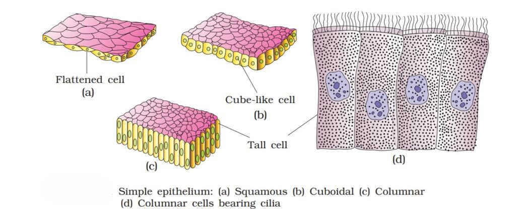
Types of Simple Epithelium (based on cell shape):
(i) Squamous Epithelium:
- Single layer of thin, flattened cells with irregular boundaries.
- Found in walls of blood vessels and air sacs of lungs.
- Function: Forms a diffusion boundary for easy exchange of substances.
(ii) Cuboidal Epithelium:
- Single layer of cube-shaped cells.
- Found in ducts of glands and tubular parts of nephrons in kidneys.
- Function: Secretion and absorption.
- Example: Proximal convoluted tubule epithelium in kidneys, often with microvilli.
(iii) Columnar Epithelium:
- Single layer of tall, slender cells with nuclei at the base.
- Free surface may have microvilli for absorption.
- Found in the lining of the stomach and intestines.
- Function: Secretion and absorption.
- Ciliated Columnar Epithelium: Columnar cells with cilia on the free surface that move particles or mucus in a specific direction. Found in bronchioles and fallopian tubes.
Question 9.
Distinguish between
- Simple epithelium and compound epithelium
- Cardiac muscle and striated muscle
- Dense regular and dense irregular connective tissues
- Adipose and blood tissue
- Simple gland and compound gland
Answer:
1. Simple epithelium and compound epithelium:
Simple epithelium is composed of a single layer of cells and mainly functions as a lining for body cavities, ducts, and tubes. In contrast, compound or stratified epithelium consists of two or more layers of cells and primarily provides protection, such as in the skin.
2. Cardiac Muscle and Striated (Skeletal) Muscle
- Cardiac Muscle:
- Found only in the heart wall.
- Short, cylindrical, branched cells with truncated ends.
- Faint striations; intercalated discs and oblique bridges present.
- Involuntary control via autonomic nervous system.
- Rich blood supply and many mitochondria; contracts rapidly without fatigue.
- Striated Muscle:
- Found in body wall, limbs, tongue, pharynx, and upper esophagus.
- Long, cylindrical, unbranched cells with blunt ends.
- Distinct striations; no intercalated discs or oblique bridges.
- Voluntary control via central nervous system.
- Rich blood supply; contracts rapidly but fatigues quickly.
3. Dense regular and dense irregular connective tissues:
In dense regular connective tissue, collagen fibers are arranged in parallel bundles, providing tensile strength in a single direction. Tendons and ligaments are examples of this tissue. Dense irregular connective tissue, however, has collagen fibers oriented in different directions, offering strength in multiple directions. This type of tissue is found in the dermis of the skin.
4. Adipose tissue and blood tissue:
Adipose tissue is a type of loose connective tissue located mainly beneath the skin. Its cells, called adipocytes, are specialized for storing fats, which serve as energy reserves, insulation, and cushioning. Blood, on the other hand, is a fluid connective tissue composed of plasma, red blood cells, white blood cells, and platelets. It acts as the main circulating fluid, transporting gases, nutrients, hormones, and metabolic wastes throughout the body.
5. Simple gland and compound gland:
Simple glands have a single, unbranched duct. They may be tubular, coiled tubular, or alveolar (acinus) in structure. Examples include the crypts of the intestine, sweat glands, and mucus-secreting glands of frog skin. Compound glands, however, have a branched duct system and may be tubular, alveolar, or tubuloalveolar. Examples include the pancreas, salivary glands, gastric glands, and functional mammary glands.
Question 10.
Mark the odd one in each series:
(a) Areolar tissue; blood; neuron; tendon
Answer:
Odd one: Neuron
Reason: Areolar tissue, blood, and tendon are connective tissues, while a neuron is a nerve tissue.
(b) RBC; WBC; platelets; cartilage
Answer:
Odd one: Cartilage
Reason: RBC, WBC, and platelets are formed elements of blood, while cartilage is a connective tissue.
(c) Exocrine; endocrine; salivary gland; ligament
Answer:
Odd one: Ligament
Reason: Exocrine, endocrine, and salivary glands are types of glands, whereas a ligament is a connective tissue.
(d) Maxilla; mandible; labrum; antennae
Answer:
Odd one: Antennae
Reason: Maxilla, mandible, and labrum are parts of the mouth in animals, whereas antennae are sensory appendages.
(e) Protonema; mesothorax; metathorax; coxa
Answer:
Odd one: Protonema
Reason: Mesothorax, metathorax, and coxa are parts of the insect body, whereas protonema is a stage in the life cycle of moss.
Question 11.
Match the terms in column I with those in column II:
Column I Column II
(a) Compound epithelium (i) Alimentary canal
(b) Compound eye (ii) Cockroach
(c) Septal nephridia (iii) Skin
(d) Open circulatory system (iv) Mosaic vision
(e) Typhlosole (v) Earthworth
(f) Osteocytes (vi) Phallomere
(Genitalia (vii) Bone
Answer:
(a) – (iii) Skin
(b) – (iv) Mosaic vision
(c) – (v) Earthworth
(d) – (ii) Cockroach
(e) – (i) Alimentary canal
(f) – (vii) Bone
(g) – (vi) Mosaic vision
Question 12.
Mention breifly about the circulatory system of earthworm
Answer:
Circulatory System of Earthworm:
- Earthworm has a closed circulatory system consisting of heart, blood vessels, and blood.
- Blood glands in the 4th, 5th, and 6th segments produce blood cells and hemoglobin, which remain dissolved in plasma.
- Pulsatile hearts are present in the 7th, 9th, 12th, and 13th segments; they pump blood in one direction.
- Smaller blood vessels supply blood to the alimentary canal, nerve cord, and body wall.
- Blood cells are phagocytic in nature, aiding in defense.
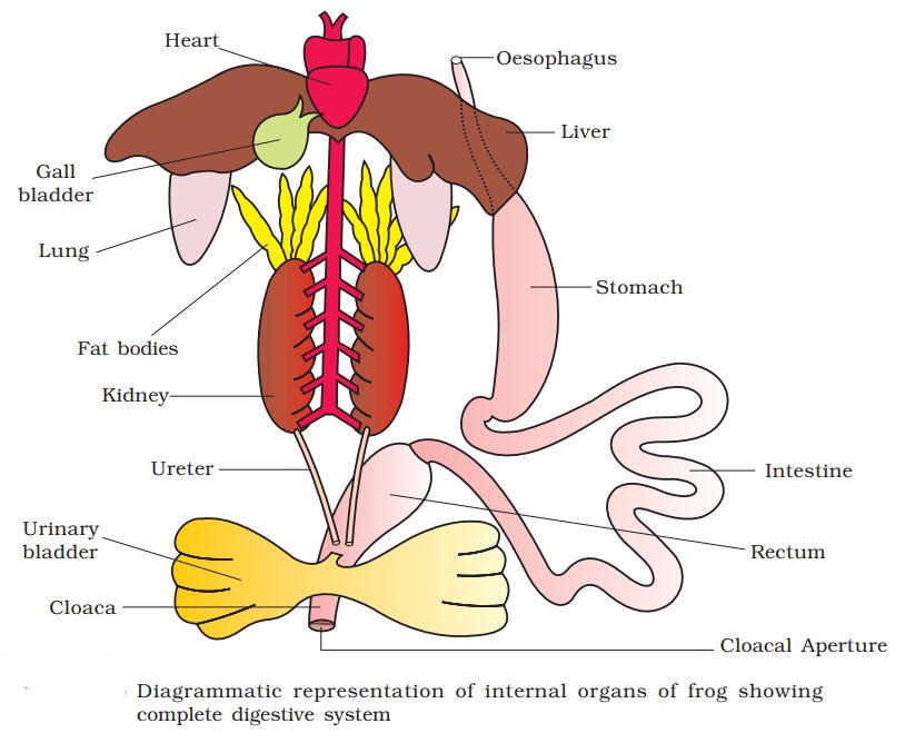
Question 13.
Draw a neat diagram of digestive system of frog.
Answer:
Question 14.
Mention the function of the following
(a) Ureters in frog
(b) Malpighian tubules
Answer:
1. Ureters in frog:
Ureters transport urine from the kidneys to the cloaca. In male frogs, they act as urinogenital ducts, while in females, ureters and oviducts open separately into the cloaca. They help excrete nitrogenous wastes (urea), making frogs ureotelic.
2. Malpighian tubules:
Malpighian tubules are thin, filamentous structures located at the junction of the midgut and hindgut in insects. They remove nitrogenous wastes from the hemolymph and help in osmoregulation.
3. Body wall in earthworm:
The body wall protects internal organs, aids in locomotion, and facilitates gas exchange. It also supports integumentary nephridia, which regulate body fluid volume and composition by collecting excess coelomic fluid and excreting it to the body surface.
Additional questions and answers
Question 1.
What is the difference between open and closed circulatory systems?
Answer:
- In an open circulatory system, blood flows freely through body cavities and is not confined to vessels (e.g., cockroach).
- In a closed circulatory system, blood flows only through vessels and does not mix with body fluids (e.g., earthworm, vertebrates).
Question 2.
Define nephridia and mention its types.
Answer:
Nephridia are excretory organs in annelids that remove nitrogenous wastes and regulate body fluids.
- Types:
- Septal nephridia (earthworm) – present on septa between segments.
- Integumentary nephridia – attached to body wall.
Question 3.
What are aortic arches in earthworm?
Answer:
Aortic arches are five pairs of pulsatile structures in segments 7, 9, 10, 11, and 13 of earthworms that connect dorsal and ventral blood vessels and act as the heart, pumping blood through the closed circulatory system.
Question 4.
Name the main components of the nervous system in earthworm.
Answer:
The nervous system of an earthworm consists of:
- A dorsal cerebral ganglion (brain)
- A ventral nerve cord with segmental ganglia
- Lateral nerves in each segment
Question 5.
What is the function of coelom in earthworm?
Answer:
The coelom is a fluid-filled cavity that acts as a hydrostatic skeleton, supports internal organs, and helps in locomotion. It also allows the distribution of nutrients and excretory products.
Question 6.
Differentiate between skeletal and smooth muscle.
Answer:
- Skeletal muscle: Voluntary, striated, multinucleated, attached to bones, fatigues quickly.
- Smooth muscle: Involuntary, unstriated, single nucleus, found in walls of hollow organs (e.g., intestine), contracts slowly and does not fatigue easily.
Question 7.
What is the function of typhlosole in earthworm?
Answer:
Typhlosole is a dorsal fold in the intestine of the earthworm that increases the surface area for absorption of nutrients.
Question 8.
Mention the types of epithelial tissue.
Answer:
- Simple epithelium: Single layer of cells; types include squamous, cuboidal, and columnar.
- Compound (stratified) epithelium: Multiple layers; provides protection.
- Ciliated epithelium: Cells bear cilia; helps in moving mucus or particles (e.g., bronchioles, fallopian tubes).