1st PUC Biology Question and Answer: Anatomy of Flowering Plants
Looking for 1st PUC Biology textbook answers? You can download Chapter 6: Anatomy of Flowering Plants Questions and Answers PDF, Notes, and Summary here. 1st PUC Biology solutions follow the Karnataka State Board Syllabus, making it easier for students to revise and score higher in exams.
Karnataka 1st PUC Biology Textbook Answers—Reflections Chapter 6
Anatomy of Flowering Plants Questions and Answers, Notes, and Summary
1st PUC Biology Chapter 6
Anatomy of Flowering Plants
Scroll Down to Download Anatomy of Flowering Plants PDF
Question and Answer:
Question 1.
State the location and function of different types of meristems.
Answer:
location and Function of Different Types of Meristems
- Apical Meristem
- Location: Found at the tips of roots and shoots.
- Function: Causes increase in length of the plant (primary growth).
- Intercalary Meristem
- Location: Present at the bases of leaves or internodes (especially in grasses).
- Function: Helps in elongation of internodes and regeneration of parts removed by grazing or mowing.
- Lateral Meristem
- Location: Found along the sides of stems and roots (e.g., vascular cambium, cork cambium).
- Function: Increases the girth (thickness) of the plant organ (secondary growth).
Question 2.
Cork cambium forms tissues that form the cork. Do you agree with this statement? Explain.
Answer
As the stem increases in girth due to vascular cambium activity, the outer cortical and epidermal layers rupture and need replacement. Hence, another meristematic tissue called cork cambium (phellogen) develops, usually in the cortex region.
- Phellogen is a thin layer of rectangular, thin-walled cells.
- It cuts off cells outwardly, which differentiate into cork (phellem).
- It cuts off cells inwardly, which form the secondary cortex (phelloderm).
Thus, cork cambium produces tissues that form the cork, providing a new protective covering.
Question 3.
Explain the process of secondary growth in the stems of woody angiosperms with the help of schematic diagrams. What is its significance?
Answer
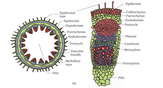
The growth of roots and stems in length with the help of apical meristem is called primary growth. In most dicotyledonous plants, there is also an increase in girth (thickness), which is known as secondary growth. The tissues responsible for secondary growth are the two lateral meristems – vascular cambium and cork cambium.
(a) Vascular Cambium
The meristematic layer that cuts off vascular tissues (xylem and phloem) is called vascular cambium. In dicot stems, the cambium present between primary xylem and primary phloem is the intrafascicular cambium. The parenchyma cells of the medullary rays adjacent to it become meristematic and form interfascicular cambium. Together, they form a continuous cambial ring.
The cambial ring becomes active and divides both towards the inner and outer sides.
- The cells formed towards the inside differentiate into secondary xylem.
- The cells formed towards the outside differentiate into secondary phloem.
Since the cambium is more active on the inner side, more secondary xylem is produced compared to phloem, forming a compact mass. The continuous formation of secondary xylem gradually crushes the primary and secondary phloem. At certain places, cambium also produces narrow bands of parenchyma, known as medullary rays, which run radially and help in lateral conduction.
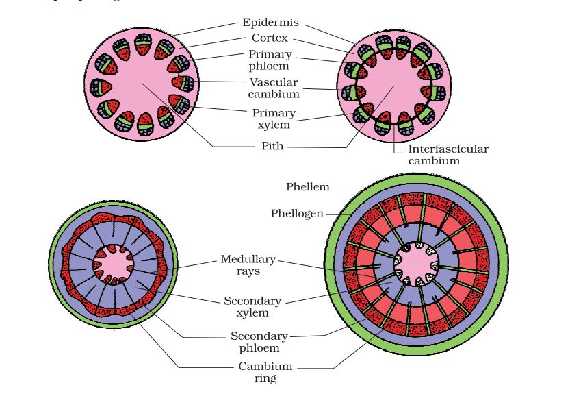
(b) Cork Cambium
As the stem increases in girth due to vascular cambium activity, the outer cortical and epidermal layers rupture. To replace them, another meristematic tissue called cork cambium (phellogen) develops in the cortex.
- The phellogen is usually a few layers thick, consisting of thin-walled rectangular cells.
- It cuts off cells on both sides:
- Outer side → cells differentiate into cork (phellem), which is suberized and impervious to water.
- Inner side → cells differentiate into secondary cortex (phelloderm), which is parenchymatous.
Together, phellogen, phellem, and phelloderm constitute the periderm. Due to cork formation, pressure builds on outer layers, which die and eventually peel off.
At some points, instead of cork, the phellogen produces loosely arranged parenchymatous cells, which rupture the epidermis and form lens-shaped openings called lenticels. Lenticels allow gaseous exchange between the atmosphere and internal tissues.
Significance of Secondary Growth
- Increases the girth of the plant, giving mechanical strength.
- Produces wood (secondary xylem), which has great economic importance.
- Leads to the formation of annual rings, useful in estimating the age of a tree.
- Cork protects the plant from mechanical injury, desiccation, and infection.
- Medullary rays and secondary tissues help in the lateral conduction of water and food.
Question 4.
Draw illustrations to bring out the anatomical difference between
Answer:
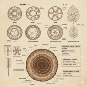
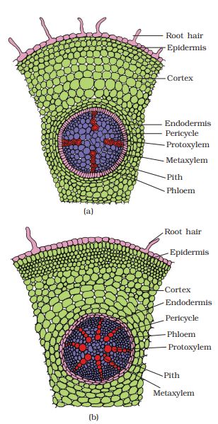
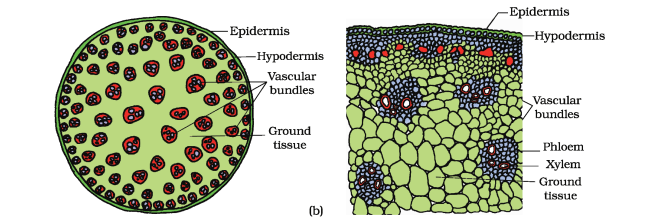
(a) Monocot root and Dicot root
(b) Monocot stem and Dicot stem
Question 5.
Cut a transverse section of young stem of a plant from your school garden and observe it under the microscope. How would you ascertain whether it is a monocot stem or a dicot stem? Answer:
Give reasons.
As shown in the above diagram the vascular bundles in dicot stems are arranged in a ring, while freely are in scattered arrangement in monocot sterns.
Question 6.
The transverse section of a plant material shows the following anatomical features
(a) the vascular bundles are conjoint, scattered and surrounded by a sclerenchyma Tous bundle sheaths.
(b) phloem parenchyma is absent. What will you identify it as?
Answer:
It is clear from the above transverse section that the plant material is a monocot stem. This is because in monocot stems:
- The vascular bundles are conjoint and scattered, each surrounded by a sclerenchymatous bundle sheath.
- Phloem parenchyma is absent, with only sieve tubes and companion cells present in phloem.
Therefore, the given transverse section belongs to a monocot stem (e.g., maize). ✅
Question 7.
Why are xylem and phloem called complex tissues?
Answer:
Xylem and phloem are called complex tissues because they are composed of several types of cells, and all these cells work together as a unit to perform common functions.
- Xylem helps in conduction of water and minerals as well as mechanical support.
- Phloem helps in the transport of food materials.
Hence, xylem and phloem are classified as complex permanent tissues. ✅
Question 8.
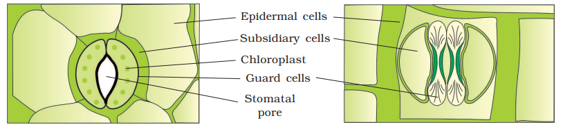
What is stomatal apparatus? Explain the structure of stomata with a labelled diagram.
Answer:
Stomata are small openings present in the epidermis of leaves. They regulate the process of transpiration and gaseous exchange.
Each stoma is composed of two bean-shaped guard cells (in dicots) or dumbbell-shaped guard cells (in grasses/monocots). The outer walls of guard cells are thin, while the inner walls are highly thickened.
The guard cells contain chloroplasts and control the opening and closing of the stomatal pore. In some cases, a few epidermal cells around the guard cells become specialized in shape and size; these are called subsidiary cells.
Thus, the stomatal apparatus includes the stomatal aperture, guard cells, and the surrounding subsidiary cells.
Question 9.
Name the three basic tissue systems in the flowering plants. Give the tissue names under each system.
Answer:
Based on their structure and location, flowering plants have three basic tissue systems:
- Epidermal Tissue System:
- Epidermis, stomata, and epidermal appendages (trichomes, root hairs)
- Ground (or Fundamental) Tissue System:
- Parenchyma, collenchyma, sclerenchyma
- Vascular (or Conducting) Tissue System:
- Xylem and phloem
Question 10.
How is the study of plant anatomy useful to us?
Answer:
The study of plant anatomy is useful in many ways. It helps us understand how plants function, including vital processes such as transpiration, photosynthesis, growth, and repair. It also aids botanists and agricultural scientists in diagnosing plant diseases and finding appropriate treatments. Moreover, since plants play a crucial role in maintaining the ecological balance of the Earth, studying their internal structure allows us to better understand the larger ecological system and the interdependence of living organisms on this planet.
Question 11.
What is periderm? How does periderm formation take place in the dicot stems?
Answer:
Periderm is a protective tissue that replaces the epidermis in dicot stems during secondary growth. As the stem increases in girth due to the activity of the vascular cambium, the outer epidermis and cortex rupture and are replaced by a new protective layer formed by the cork cambium (phellogen), which usually develops in the cortex.
Phellogen is a few layers thick and cuts off cells on both sides: the outer cells differentiate into cork (phellem), which is dead and impermeable to water due to suberin, while the inner cells differentiate into secondary cortex (phelloderm), which is parenchymatous. Together, the phellem, phellogen, and phelloderm constitute the periderm. Pressure from cork formation eventually causes the outer layers to die and slough off.
Question 12.
Describe the internal structure of a dorsiventral leaf with the help of labelled diagrams.
Answer:
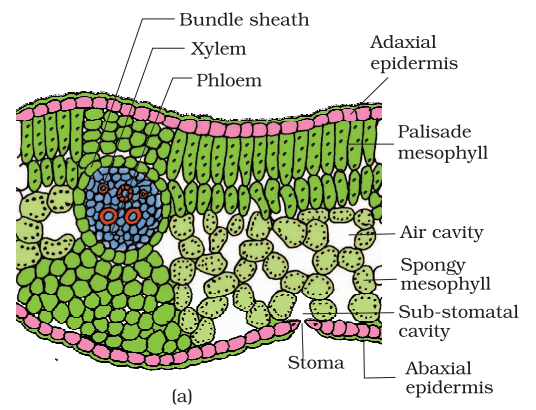
The vertical section of a dorsiventral (dicot) leaf shows three main parts: epidermis, mesophyll, and vascular system. The epidermis covers both the upper (adaxial) and lower (abaxial) surfaces of the leaf and is often coated with a conspicuous cuticle. The abaxial epidermis usually has more stomata than the adaxial epidermis, which may sometimes lack stomata. The tissue between the two epidermal layers is the mesophyll, which contains chloroplasts and performs photosynthesis.
The mesophyll is differentiated into palisade parenchyma (elongated, vertically arranged cells just below the adaxial epidermis) and spongy parenchyma (loosely arranged oval or round cells below the palisade layer with large intercellular spaces for gaseous exchange).
The vascular system is present in the veins and midrib, with the size of vascular bundles varying according to the thickness of the veins. Each vascular bundle is surrounded by a layer of thick-walled bundle sheath cells, providing support and protection. The arrangement of veins shows reticulate venation, characteristic of dicot leaves.