1st PUC Biology Question and Answer: Neural Control And Coordination
Looking for 1st PUC Biology textbook answers? You can download Chapter 21: Neural Control And Coordination Questions and Answers PDF, Notes, and Summary here. 1st PUC Biology solutions follow the Karnataka State Board Syllabus, making it easier for students to revise and score higher in exams.
Karnataka 1st PUC Biology Textbook Answers—Reflections Chapter 21
Neural Control And Coordination Questions and Answers, Notes, and Summary
1st PUC Biology Chapter 21
Neural Control And Coordination

Scroll Down to Downlaod Neural Control And Coordination PDF
Question and Answer:
Question 1.
Briefly describe the structure of the following:
(a) Brain
(b) Eye
(c) Ear
Answer:
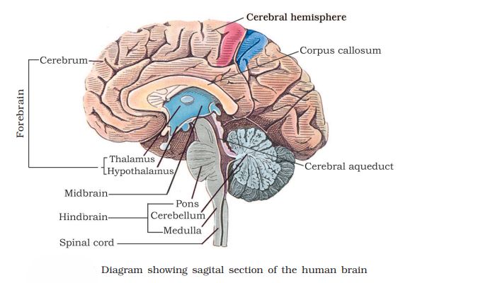
(a) Brain:
The human brain is protected by the skull and covered by three meninges – dura mater, arachnoid, and pia mater. It is divided into the forebrain, midbrain, and hindbrain. The forebrain includes the cerebrum, thalamus, and hypothalamus. The cerebrum has two hemispheres and is responsible for intelligence, memory, and sensory activities.
The thalamus relays sensory and motor signals, while the hypothalamus controls hunger, thirst, and body temperature. The midbrain connects the forebrain and hindbrain and controls reflexes. The hindbrain consists of the pons, cerebellum, and medulla oblongata, which regulate balance and involuntary actions such as heartbeat and breathing.
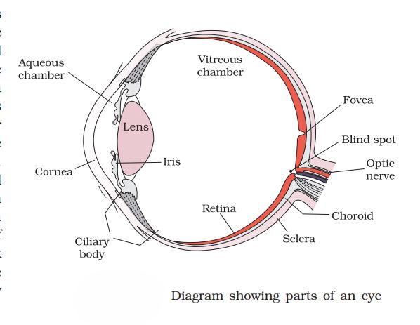
(b) Eye:
The human eye is a spherical structure with three layers – sclera, choroid, and retina. The outer sclera protects the eye, the choroid is rich in blood vessels and forms the ciliary body and iris, and the retina contains photoreceptor cells (rods and cones). The pupil, controlled by the iris, regulates light entry.
The transparent lens focuses light on the retina. The aqueous humor (in front of the lens) and vitreous humor (behind the lens) maintain the eye’s shape. The optic nerve carries visual impulses to the brain, while the fovea provides the sharpest vision.
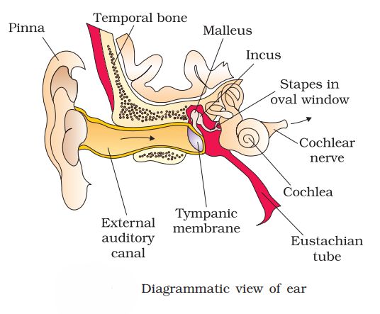
(c) Ear:
The ear has three parts – outer, middle, and inner ear. The outer ear includes the pinna and ear canal, ending in the eardrum. The middle ear has three small bones – malleus, incus, and stapes – which transmit sound vibrations to the inner ear. The Eustachian tube connects the middle ear to the pharynx and balances air pressure.
The inner ear consists of the cochlea and vestibular apparatus. The cochlea contains the organ of Corti for hearing, while the vestibular apparatus (semicircular canals, saccule, and utricle) helps maintain balance.
Question 2.
Compare the following:
- Central neural system (CNS) and Peripheral neural system (PNS)
- Resting potential and action potential
- Choroid and retina
Answer:
(a) Central Neural System (CNS) and Peripheral Neural System (PNS):
The central neural system consists of the brain and spinal cord, which act as the main control centre for processing information and coordinating body activities. The peripheral neural system includes all the nerves that arise from the brain and spinal cord, connecting the CNS to different parts of the body. While the CNS interprets and processes information, the PNS transmits signals between the CNS and the rest of the body.
(b) Resting Potential and Action Potential:
When a neuron is not transmitting any impulse, the membrane shows resting potential, with the inside of the neuron being negatively charged compared to the outside due to more sodium ions outside and potassium ions inside. During the transmission of an impulse, the membrane becomes depolarized, and the inside becomes positively charged — this change is called the action potential. Thus, resting potential represents the inactive state, while action potential represents the active transmission of a nerve impulse.
(c) Choroid and Retina:
The choroid is the middle, dark, and vascular layer of the eyeball that provides nourishment to the eye tissues. It forms the ciliary body and iris in the front part of the eye. The retina is the innermost sensory layer that contains photoreceptor cells — rods and cones — which detect light and colour and convert them into nerve impulses. Hence, the choroid mainly nourishes the eye, while the retina is responsible for vision.
Question 3.
Explain the following processes:
(a) Polarisation of the membrane of a nerve fibre
(b) Depolarisation of the membrane of a nerve fibre
(c) Conduction of a nerve impulse along a nerve fibre
(d) Transmission of a nerve impulse across a chemical synapse
Answer:
(a) Polarisation of the membrane of a nerve fibre:
When a neuron is at rest, its membrane is said to be polarised. The axon membrane is more permeable to potassium ions (K⁺) and almost impermeable to sodium ions (Na⁺) and negatively charged proteins. As a result, the axoplasm has a high concentration of K⁺ and negatively charged proteins, while the fluid outside contains more Na⁺.
This difference is maintained by the sodium-potassium pump, which actively transports three Na⁺ ions out and two K⁺ ions in. Hence, the outer surface becomes positively charged and the inner surface negatively charged, creating the resting potential across the membrane.
(b) Depolarisation of the membrane of a nerve fibre:
When a stimulus is applied, the permeability of the membrane changes, allowing sodium ions to rush inside the neuron. This causes the inner surface to become positively charged and the outer surface negative, leading to depolarisation. The electrical potential that appears during this phase is called the action potential, which represents the nerve impulse.
(c) Conduction of a nerve impulse along a nerve fibre:
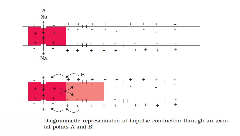
When depolarisation occurs at one point on the axon, it triggers a local current flow that causes the adjacent region of the membrane to also depolarise. The area behind it then returns to its resting state. This sequence of depolarisation and repolarisation continues along the length of the axon, resulting in the conduction of the nerve impulse from one end to the other.
(d) Transmission of a nerve impulse across a chemical synapse:
At a synapse, the axon terminal of one neuron is separated from the next neuron by a small gap called the synaptic cleft. When an impulse reaches the axon terminal, it stimulates synaptic vesicles to release neurotransmitters into the cleft. These chemicals bind to receptors on the postsynaptic membrane, opening ion channels and allowing ions to enter. This generates a new potential in the next neuron, which may be excitatory or inhibitory, thus completing the transmission of the nerve impulse.
Question 4.
Draw labelled diagrams of the following:
- Neuron
- Brain
- Eye
- Ear
Answer:
(a) Neuron:
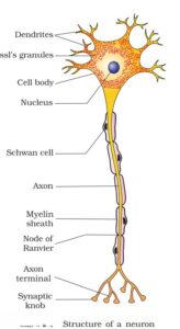
A neuron is the structural and functional unit of the nervous system. It consists of a cell body, dendrites, and an axon. The cell body contains the nucleus and cytoplasm. Dendrites receive impulses, while the axon carries impulses away from the cell body.
(Draw a neat labelled diagram of a neuron.)
(b) Brain:
The human brain is protected by the skull and covered by three meninges — dura mater, arachnoid, and pia mater. It is divided into forebrain, midbrain, and hindbrain.
- Forebrain: Includes cerebrum, thalamus, and hypothalamus; controls memory, reasoning, and sensory functions.
- Midbrain: Connects the forebrain and hindbrain; has corpora quadrigemina for reflexes.
- Hindbrain: Includes pons, cerebellum, and medulla; controls balance and vital activities.
(Draw a neat labelled diagram of the human brain.)
(c) Eye:
The human eye is a spherical organ with three layers — sclera, choroid, and retina. It contains structures like cornea, iris, pupil, lens, and retina. The retina has photoreceptors — rods (for dim light) and cones (for colour vision). Fovea is the point of sharpest vision and blind spot lacks photoreceptors.
(Draw a neat labelled diagram of the human eye.)
(d) Ear:
The ear has three parts — outer ear, middle ear, and inner ear.
- Outer ear: Includes pinna and auditory canal.
- Middle ear: Has three ossicles — malleus, incus, and stapes — that transmit sound vibrations.
- Inner ear: Consists of cochlea (for hearing) and semicircular canals (for balance).
(Draw a neat labelled diagram of the human ear.)
Question 5.
Write short notes on the following:
- Neural coordination
- Forebrain
- Midbrain
- Hindbrain
- Retina
- Ear ossicles
- Cochlea
- Organ of Corti
- Synapse
Answer:
(a) Neural coordination:
Neural coordination is the process through which the nervous system controls and coordinates the activities of different parts of the body. It involves the reception of stimuli, transmission of nerve impulses, processing of information by the brain and spinal cord, and sending appropriate responses to effectors like muscles and glands.
(b) Forebrain:
The forebrain consists of the cerebrum, thalamus, and hypothalamus. The cerebrum is the largest part, responsible for intelligence, memory, reasoning, and voluntary actions. The thalamus acts as a relay center for sensory and motor signals, while the hypothalamus regulates body temperature, hunger, thirst, and emotions.
(c) Midbrain:
The midbrain lies between the forebrain and the hindbrain. It contains the cerebral aqueduct and four swellings called corpora quadrigemina. The midbrain controls reflexes related to vision and hearing and helps in the transmission of motor signals.
(d) Hindbrain:
The hindbrain includes the pons, cerebellum, and medulla oblongata. The pons connects different parts of the brain, the cerebellum maintains balance and posture, and the medulla controls involuntary activities like heartbeat, breathing, and blood pressure.
(e) Retina:
The retina is the innermost layer of the eye containing photoreceptor cells—rods and cones. Rods help in vision in dim light, while cones are responsible for colour vision. The retina also contains the blind spot (no photoreceptors) and the fovea (region of sharp vision).
(f) Ear ossicles:
The ear ossicles are three tiny bones in the middle ear—malleus, incus, and stapes. They are arranged in a chain and help in transmitting and amplifying sound vibrations from the tympanic membrane to the oval window of the cochlea.
(g) Cochlea:
The cochlea is a coiled, spiral-shaped structure in the inner ear. It contains fluid-filled chambers and the organ of Corti. The cochlea converts sound vibrations into nerve impulses which are transmitted to the brain for sound perception.
(h) Organ of Corti:
The organ of Corti is located on the basilar membrane inside the cochlea. It contains hair cells that act as auditory receptors. Vibrations in the cochlear fluid move these hair cells, generating nerve impulses sent to the auditory nerve.
(i) Synapse:
A synapse is a junction between two neurons where nerve impulses are transmitted. In a chemical synapse, neurotransmitters released from the presynaptic neuron cross the synaptic cleft and bind to receptors on the postsynaptic neuron, generating a new nerve impulse.
Question 6.
Give a brief account of:
(a) Mechanism of synaptic transmission
(b) Mechanism of vision
(c) Mechanism of hearing
Answer:
(a) Mechanism of synaptic transmission:
The transmission of nerve impulses across a synapse occurs through chemical messengers called neurotransmitters. When an impulse reaches the axon terminal of the presynaptic neuron, it triggers the release of neurotransmitters (such as acetylcholine) from synaptic vesicles into the synaptic cleft. These neurotransmitters bind to specific receptors on the postsynaptic membrane, causing ion channels to open and generating a new action potential in the postsynaptic neuron. The neurotransmitters are then broken down or reabsorbed, ending the signal transmission.
(b) Mechanism of vision:
The light rays from an object enter the eye through the cornea and pass through the aqueous humor, pupil, lens, and vitreous humor before reaching the retina. The lens focuses the light on the retina, where photoreceptor cells—rods and cones—convert the light energy into electrical impulses. These impulses are transmitted via the optic nerve to the visual cortex of the brain, where the image is interpreted. Rods are responsible for vision in dim light, while cones help in colour vision and sharpness.
(c) Mechanism of hearing:
Sound waves are collected by the pinna and directed through the auditory canal to strike the tympanic membrane (eardrum), causing it to vibrate. These vibrations are transmitted and amplified by the three ear ossicles—malleus, incus, and stapes—to the oval window of the cochlea. The movement of the oval window sets the fluid in the cochlea into motion, stimulating the hair cells of the organ of Corti. These hair cells convert sound vibrations into nerve impulses, which are carried by the auditory nerve to the brain, where the sound is perceived and interpreted.
Question 7.
Answer briefly:
(a) How do you perceive the colour of an object?
(b) Which part of our body helps us in maintaining the body balance?
(c) How does the eye regulate the amount of light that falls on the retina.
Answer:
(a) How do you perceive the colour of an object?
The colour of an object is perceived by the cone cells present in the retina of the eye. There are three types of cone cells, each sensitive to red, green, or blue light. When light reflected from an object enters the eye, different cones are stimulated in varying degrees, and the combination of signals is interpreted by the brain as different colours.
(b) Which part of our body helps us in maintaining the body balance?
The vestibular apparatus located in the inner ear helps in maintaining body balance. It consists of three semicircular canals and two otolith organs (utricle and saccule) that detect changes in head position and movement, helping the body maintain equilibrium.
(c) How does the eye regulate the amount of light that falls on the retina?
The iris controls the size of the pupil, thereby regulating the amount of light entering the eye. In bright light, the pupil constricts to reduce light entry, while in dim light, the pupil dilates to allow more light to reach the retina.
Question 8.
Explain the following:
(a) Role of Na in the generation of action potential.
(b) Mechanism of generation of light-induced impulse in the retina.
(c) Mechanism through which a sound produces a nerve impulse in the inner ear.
Answer:
(a) Role of Na⁺ in the generation of action potential:
When a neuron is at rest, the inside of the axonal membrane is negatively charged compared to the outside due to the unequal distribution of ions. When a stimulus is applied, the permeability of the membrane to sodium ions (Na⁺) increases suddenly. Na⁺ ions rapidly move into the neuron, causing the inner side of the membrane to become positively charged. This reversal of polarity is called depolarization, and it generates an action potential, which is the basis for nerve impulse transmission.
(b) Mechanism of generation of light-induced impulse in the retina:
In the retina, photoreceptor cells (rods and cones) contain light-sensitive pigments. In rods, the pigment is rhodopsin (formed from opsin and retinal). When light strikes rhodopsin, it splits into opsin and retinal, leading to a change in membrane potential (hyperpolarization). This change triggers electrical impulses in the photoreceptor cells, which are then transmitted through bipolar and ganglion cells to form nerve impulses. These impulses travel via the optic nerve to the brain, where the visual image is interpreted.
(c) Mechanism through which a sound produces a nerve impulse in the inner ear:
Sound waves set the tympanic membrane (eardrum) into vibration, which are transmitted through the ear ossicles (malleus, incus, and stapes) to the oval window of the cochlea. The movement of the oval window creates pressure waves in the cochlear fluid, causing the basilar membrane to vibrate. These vibrations bend the hair cells of the organ of Corti against the tectorial membrane, generating electrical impulses. These impulses are then transmitted by the auditory nerve to the brain, where the sound is perceived.
Question 9.
Differentiate between:
(a) Myelinated and non-myelinated axons
(b) Dendrites and axons
(c) Rods and cones
(d) Thalamus and Hypothalamus
(e) Cerebrum and Cerebellum
Answer:
(a) Difference between Myelinated and Non-myelinated Axons
Feature | Myelinated Axon | Non-myelinated Axon |
1. Myelin sheath | Covered by a myelin sheath made of Schwann cells. | Lacks a myelin sheath. |
2. Speed of impulse | Conducts nerve impulses rapidly by saltatory conduction. | Conducts nerve impulses slowly. |
3. Nodes of Ranvier | Present between two myelinated segments. | Absent. |
4. Location | Found in the white matter of the brain and spinal cord. | Found in the grey matter and autonomic nerves. |
(b) Difference between Dendrites and Axons
Feature | Dendrites | Axons |
1. Function | Receive impulses from other neurons. | Transmit impulses away from the cell body. |
2. Number | Usually short and many. | Single and long. |
3. Direction of impulse | Carry impulse towards the cell body. | Carry impulse away from the cell body. |
4. Branching | Highly branched near the cell body. | Branches only at the terminal end. |
(c) Difference between Rods and Cones
Feature | Rods | Cones |
1. Function | Responsible for vision in dim light (night vision). | Responsible for vision in bright light and colour vision. |
2. Pigment | Contain the pigment rhodopsin. | Contain the pigment iodopsin. |
3. Sensitivity | Highly sensitive to light but not to colour. | Sensitive to colour but less sensitive to light. |
4. Distribution | More numerous around the periphery of retina. | Concentrated in the fovea centralis. |
(d) Difference between Thalamus and Hypothalamus
Feature | Thalamus | Hypothalamus |
1. Location | Located above the hypothalamus. | Located below the thalamus. |
2. Function | Acts as a relay centre for sensory and motor signals to the cerebrum. | Regulates body temperature, hunger, thirst, sleep, and hormone secretion. |
3. Endocrine role | No direct endocrine function. | Contains neurosecretory cells that control the pituitary gland. |
(e) Difference between Cerebrum and Cerebellum
Feature | Cerebrum | Cerebellum |
1. Location | Largest part of the forebrain. | Part of the hindbrain located below the cerebrum. |
2. Function | Controls intelligence, memory, learning, and voluntary actions. | Maintains body posture, balance, and coordination of movements. |
3. Structure | Divided into two hemispheres with many folds. | Smaller, highly folded structure with two hemispheres. |
4. Type of control | Controls voluntary activities. | Controls involuntary muscular coordination. |
Question 10.
Answer the following:
(a) Which part of the ear determines the pitch of a sound?
(b) Which part of the human brain is the most developed?
(c) Which part of our central neural system acts as a master clock?
Answer:
(a) Organ of Corti – It is the part of the cochlea in the inner ear that helps determine the pitch of a sound by responding to different sound frequencies.
(b) Cerebrum – It is the most developed part of the human brain, responsible for intelligence, reasoning, memory, learning, and voluntary actions.
(c) Pineal gland – It acts as the master clock of our central neural system by regulating circadian rhythms (sleep–wake cycle) through the secretion of the hormone melatonin.
Question 11.
The region of the vertebrate eye, where the optic nerve passes out of the retina, is called the
(a) fovea
(b) iris
(c) blind spot
(d) optic chaisma
✅ (c) Blind spot
Explanation:
The blind spot is the region of the retina where the optic nerve exits the eye. This area lacks photoreceptor cells (rods and cones), so no visual image is formed there, hence it is called the blind spot.
Question 12.
Distinguish between:
(a) afferent neurons and efferent neurons
(b) impulse conduction in a myelinated nerve fibre and unmyelinated nerve fibre
(c) aqueous humor and vitreous humor
(d) blind spot and yellow spot
(f) cranial nerves and spinal nerves.
Answer:
(a) Difference between Afferent Neurons and Efferent Neurons
Feature | Afferent Neurons | Efferent Neurons |
1. Function | Carry impulses from sense organs to the central nervous system (CNS). | Carry impulses from CNS to effectors (muscles or glands). |
2. Direction of impulse | Towards the CNS. | Away from the CNS. |
3. Also called | Sensory neurons. | Motor neurons. |
4. Example | Neurons connected to eyes, ears, and skin. | Neurons controlling muscles and glands. |
(b) Difference between Impulse Conduction in Myelinated and Unmyelinated Nerve Fibres
Feature | Myelinated Nerve Fibre | Unmyelinated Nerve Fibre |
1. Myelin sheath | Present around the axon. | Absent. |
2. Mode of conduction | Impulse jumps from one node of Ranvier to the next (saltatory conduction). | Impulse travels continuously along the fibre. |
3. Speed of conduction | Very fast. | Slow. |
4. Energy usage | Requires less energy. | Requires more energy. |
(c) Difference between Aqueous Humor and Vitreous Humor
Feature | Aqueous Humor | Vitreous Humor |
1. Location | Between cornea and lens (aqueous chamber). | Between lens and retina (vitreous chamber). |
2. Nature | Clear, watery fluid. | Transparent, gel-like fluid. |
3. Function | Maintains intraocular pressure and nourishes the lens and cornea. | Maintains the shape of the eyeball and supports the retina. |
(d) Difference between Blind Spot and Yellow Spot
Feature | Blind Spot | Yellow Spot (Macula Lutea) |
1. Photoreceptors | Absent (no rods or cones). | Only cones are present. |
2. Function | No image formation occurs here. | Region of maximum visual acuity. |
3. Location | Point where optic nerve leaves the retina. | Lateral to the blind spot. |
(f) Difference between Cranial Nerves and Spinal Nerves
Feature | Cranial Nerves | Spinal Nerves |
1. Origin | Arise from the brain. | Arise from the spinal cord. |
2. Number | 12 pairs. | 31 pairs. |
3. Function | Connect brain with head and neck organs. | Connect spinal cord with body parts below the neck. |
4. Type | May be sensory, motor, or mixed. | All are mixed nerves. |
Additional Questions and Answers
Question 1.
What is the structural and functional unit of the nervous system?
Answer:
The neuron is the structural and functional unit of the nervous system. It is specialized for the transmission of nerve impulses.
Question 2.
Name the three main parts of a neuron and mention their functions.
Answer:
- Cell body (Cyton): Contains the nucleus and cytoplasm; controls metabolic activities.
- Dendrites: Receive impulses from other neurons.
- Axon: Conducts impulses away from the cell body.
Question 3.
What is a synapse?
Answer:
A synapse is a functional junction between two neurons, where the transmission of nerve impulse occurs with the help of neurotransmitters like acetylcholine.
Question 4.
What are meninges? Name their layers.
Answer:
Meninges are protective membranes that cover the brain and spinal cord.
They consist of three layers:
- Dura mater (outermost tough layer)
- Arachnoid mater (middle web-like layer)
- Pia mater (innermost delicate layer)
Question 5.
What is cerebrospinal fluid (CSF)? State its function.
Answer:
Cerebrospinal fluid is a clear fluid that surrounds the brain and spinal cord.
Functions:
- Protects the brain from shocks.
- Maintains constant pressure around the brain.
- Helps in exchange of nutrients and waste products.
Question 6.
What are the functions of the medulla oblongata?
Answer:
The medulla oblongata controls involuntary functions such as heartbeat, breathing, blood pressure, and swallowing.
Question 7.
Define reflex action. Give an example.
Answer:
Reflex action is an automatic, involuntary, and immediate response to a stimulus.
Example: Withdrawal of hand when touching a hot object.
Question 8.
What is a reflex arc?
Answer:
A reflex arc is the pathway followed by nerve impulses during a reflex action. It includes receptor, sensory neuron, interneuron, motor neuron, and effector.
Question 9.
What are rods and cones? Mention their functions.
Answer:
- Rods: Photoreceptor cells sensitive to dim light; help in night vision.
- Cones: Photoreceptor cells sensitive to bright light and color; help in color vision.
Question 10.
What is the function of the semicircular canals in the ear?
Answer:
Semicircular canals help in maintaining body balance and equilibrium.
Question 11.
Name the parts of the human ear.
Answer:
The ear has three parts:
- Outer ear – pinna and auditory canal
- Middle ear – tympanum and ossicles (malleus, incus, stapes)
- Inner ear – cochlea and semicircular canals
Question 12.
What is the function of the cerebellum?
Answer:
The cerebellum coordinates voluntary movements and maintains balance, posture, and muscle tone.
Question 13.
What is saltatory conduction?
Answer:
Saltatory conduction is the jumping of nerve impulses from one node of Ranvier to the next in a myelinated nerve fibre, increasing the speed of transmission.
Question 14.
What is the function of the hypothalamus?
Answer:
The hypothalamus regulates body temperature, hunger, thirst, emotions, and secretion of pituitary hormones.
Question 15.
Name the three ossicles of the middle ear and their functions.
Answer:
The three ossicles are:
- Malleus
- Incus
- Stapes
They amplify and transmit sound vibrations from the tympanic membrane to the inner ear.