1st PUC Biology Question and Answer: Locomotion And Movement
Looking for 1st PUC Biology textbook answers? You can download Chapter 20: Locomotion And Movement Questions and Answers PDF, Notes, and Summary here. 1st PUC Biology solutions follow the Karnataka State Board Syllabus, making it easier for students to revise and score higher in exams.
Karnataka 1st PUC Biology Textbook Answers—Reflections Chapter 20
Locomotion And Movement Questions and Answers, Notes, and Summary
1st PUC Biology Chapter 20
Locomotion And Movement
Scroll Down to Download Locomotion And Movement PDF
Question and Answer.
Question 1.
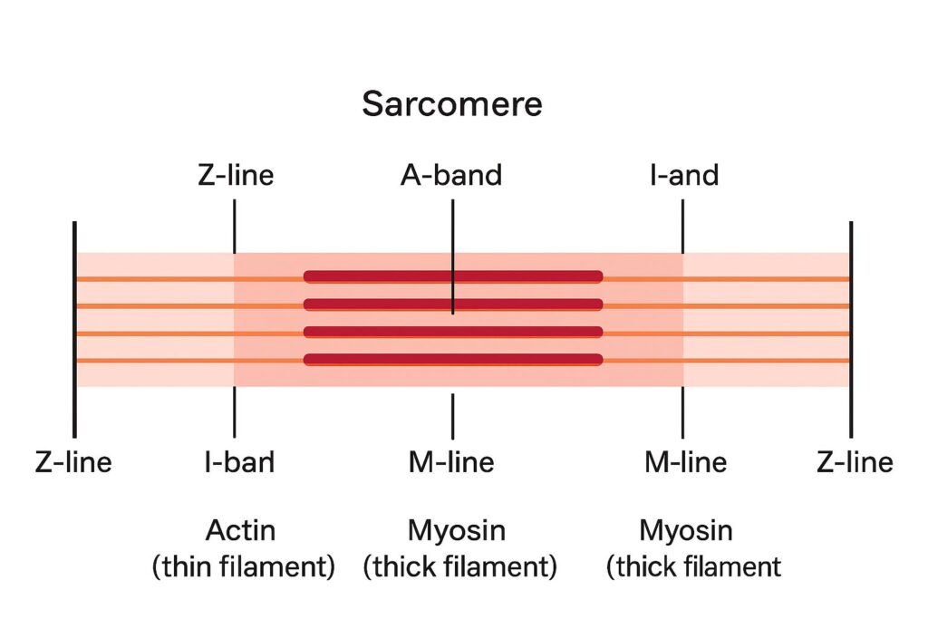
Draw the diagram of a sarcomere of skeletal muscle showing different regions.
Question 2.
Define sliding filament theory of muscle contraction.
Answer:
The sliding filament theory states that muscle contraction occurs when the thin filaments (actin) slide over the thick filaments (myosin), resulting in the shortening of the sarcomere, which ultimately leads to the contraction of the muscle fiber.
Question 3.
Describe the important steps in muscle contraction.
Answer:
Steps in Muscle Contraction:
- A nerve impulse arriving at the neuromuscular junction initiates the contractile response.
- A neurotransmitter released at the junction enters the sarcomere through its membrane channels.
- The opening of these channels allows Na⁺ ions to flow into the sarcomere, generating an action potential that travels along the entire muscle fiber.
- The sarcoplasmic reticulum releases Ca²⁺ ions, which bind to troponin on the thin filament.
- This binding causes conformational changes in troponin, exposing the active sites on F-actin.
- The myosin heads attach to these active sites, forming cross-bridges.
- The cross-bridges pull the actin filaments past the myosin filaments, shortening the sarcomere.
- When thousands of actin and myosin filaments interact simultaneously, the entire muscle cell contracts.
This process is explained by the sliding filament theory of muscle contraction.
Question 4.
Write true or false. If false change the statement so that it is true.
(a) Actin is present in thin filament
(b) H-zone of striated muscle fibre represents both thick and thin filaments.
(c) Human skeleton has 206 bones.
(d) There are 11 pairs of ribs in man.
(e) Sternum is present on the ventral side of the body.
Answer:
(a) True – Actin is present in thin filaments.
(b) False – Correct statement: H-zone of a striated muscle fibre contains only thick filaments.
(c) True – Human skeleton has 206 bones.
(d) False – Correct statement: There are 12 pairs of ribs in man.
(e) True – Sternum is present on the ventral side of the body.
Question 5.
Write the difference between:
(a) Actin and Myosin
(b) Red and White muscles
(c) Pectoral and Pelvic girdle
Answer:
(a) Actin and Myosin
- Actin Filaments:
- Thinner compared to myosin filaments.
- Exist in two forms: Globular actin (G-actin) and Fibrous actin (F-actin).
- Contain proteins tropomyosin and troponin.
- Myosin Filaments:
- Composed of two portions: Head and Tail.
- Head is formed of Heavy Meromyosin (HMM) and tail of Light Meromyosin (LMM).
- Myosin head has contractile property and ATPase activity, enabling muscle contraction.
(b) Red and White Muscles
Feature | Red Muscle | White Muscle |
Colour | Red due to high myoglobin | White due to low myoglobin |
Thickness | Thin | Thick |
Contraction | Slow and sustained | Fast |
Energy Source | Aerobic metabolism | Anaerobic metabolism |
Mitochondria | Large amount | Small amount |
Function | Perform sustained work for long duration | Perform strenuous work for short duration |
Blood Supply | Many blood capillaries (e.g., extensor muscles of back) | Fewer blood capillaries (e.g., muscles of eyeball) |
(c) Pectoral and Pelvic Girdle
Feature | Pectoral Girdle | Pelvic Girdle |
Function | Connects forelimb with axial skeleton | Connects leg with axial skeleton |
Bones | Two bones: Clavicle and Scapula | Three bones: Ilium, Ischium, Pubis |
Fusion | Two halves are not fused | Two halves are fused |
Location | Found on backside of thorax | Found in hip region |
Socket | Glenoid cavity | Acetabulum |
Question 6.
Match Column I with Column II:
Column I Column II
(a) Smooth muscle (i) Myoglobin
(b) Tropomyosin (ii) Thin filament
(c) Red muscle (iii) Sutures
(d) Skull (iv) Involuntary
Answer:
Column I | Column II |
(a) Smooth muscle | (iv) Involuntary |
(b) Tropomyosin | (ii) Thin filament |
(c) Red muscle | (i) Myoglobin |
(d) Skull | (iii) Sutures |
Question 7.
What are the different types of movements exhibited by the cells of human body?
Answer:
Cells of the human body exhibit three main types of movements: amoeboid, ciliary, and muscular.
- Amoeboid Movement:
- Seen in specialized cells like macrophages and leukocytes in blood.
- Movement occurs by pseudopodia formed through the streaming of protoplasm (similar to Amoeba).
- Cytoskeletal elements, such as microfilaments, play an important role in this type of movement.
- Ciliary Movement:
- Occurs in internal tubular organs lined by ciliated epithelium.
- Coordinated ciliary movements help remove dust and foreign particles from the trachea.
- Also facilitates the passage of ova through the female reproductive tract.
- Muscular Movement:
- Responsible for movements of limbs, jaws, tongue, and other body parts.
- Achieved through the contractile property of muscles.
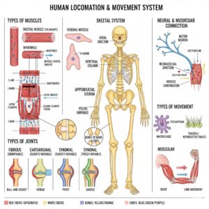
- Locomotion requires coordinated activity of the muscular, skeletal, and neural systems
I can also make a simple diagram showing amoeboid, ciliary, and muscular movements to visually explain
Question 8.
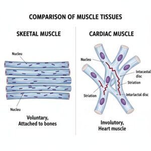
How do you distinguish between a skeletal muscle and a cardiac muscle?
Answer:
Skeletal muscle: Skeletal muscles are closely associated with the skeletal components of the body. They have a striped appearance under the microscope and hence are called striated muscles. As their activities are under the voluntary control of the nervous system, they are known as voluntary muscles. They are primarily involved in locomotory actions and changes in body posture.
Cardiac muscle: Cardiac muscles are the muscles of the heart. Many cardiac muscle cells assemble in a branching pattern to form a cardiac muscle. Based on appearance, cardiac muscles are striated. They are involuntary in nature, as the nervous system does not control their activities directly.
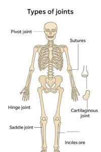
Question 9.
Name the type of joint between the following:-
(a) atlas/axis
(b) carpal/metacarpal of thumb
(c) between phalanges
(d) femur/acetabulum
(e) between cranial bones
(f) between pubic bones in the pelvic girdle
Answer:
(a) Atlas/Axis – Pivot joint (allows rotational movement of the head)
(b) Carpal/Metacarpal of thumb – Saddle joint (allows flexion, extension, abduction, adduction, and circumduction)
(c) Between phalanges – Hinge joint (allows flexion and extension)
(d) Femur/Acetabulum – Ball and socket joint (allows movement in all directions)
(e) Between cranial bones – Immovable or Fibrous joint (Sutures)
(f) Between pubic bones in the pelvic girdle – Cartilaginous joint (Pubic symphysis)
Question 10.
Fill in the blank spaces:
(a) All mammals (except a few) have cervical vertebra.
(b) The number of phalanges in each limb of human is
(c) Thin filament of myofibril contains 2 ‘F’ actins and two other proteins namely and
(d) In a muscle fibre Ca” is stored in
(e) and pairs of ribs are called floating ribs.
(f) The human cranium is made of bones.
Answer:
(a) All mammals (except a few) have 7 cervical vertebrae.
(b) The number of phalanges in each limb of human is 14.
(c) Thin filament of myofibril contains 2 F-actins and two other proteins namely troponin and tropomyosin.
(d) In a muscle fibre, Ca²⁺ is stored in sarcoplasmic reticulum.
(e) 11th and 12th pairs of ribs are called floating ribs.
(f) The human cranium is made of 8 bones.
Additional Questions & Answers
Question 1.
Define locomotion. Give examples.
Answer:
Locomotion is a voluntary movement that results in a change of place or location in an organism. Examples include walking, running, swimming, flying, and climbing.
Question 2.
Explain the difference between movement and locomotion.
Answer:
All locomotions are movements, but all movements are not locomotions. Movement may not involve change in location (e.g., eyelid blinking), whereas locomotion involves change in place (e.g., walking, swimming).
Question 3.
Name three types of cellular movements in humans and give an example of each.
Answer:
- Amoeboid movement – by pseudopodia (e.g., macrophages).
- Ciliary movement – by cilia (e.g., passage of ova in female reproductive tract).
- Muscular movement – by muscle contraction (e.g., limb movement).
Question 4.
What are the special properties of muscles?
Answer:
Muscles have four special properties:
- Excitability
- Contractility
- Extensibility
- Elasticity
Question 5.
Differentiate between skeletal, visceral, and cardiac muscles.
Answer:
Feature | Skeletal Muscle | Visceral Muscle | Cardiac Muscle |
Location | Attached to bones | Walls of hollow organs | Heart |
Appearance | Striated | Smooth | Striated & branched |
Control | Voluntary | Involuntary | Involuntary |
Function | Locomotion & posture | Transport of substances | Pumping blood |
Question 6.
What is a sarcomere?
Answer:
A sarcomere is the functional unit of a myofibril in a skeletal muscle, extending from one ‘Z’ line to the next, responsible for muscle contraction.
Question 7.
Explain the structure of actin and myosin filaments.
Answer:
- Actin (thin) filament: Made of two F-actins helically wound; associated with tropomyosin and troponin. Active sites for myosin are masked by troponin in the resting state.
- Myosin (thick) filament: Made of ~300 myosin molecules, each having a globular head (HMM) and a tail (LMM). The head has ATPase activity and binding sites for actin.
Question 8.
Describe the sliding filament theory of muscle contraction.
Answer:
Contraction occurs by sliding of actin filaments over myosin filaments. Ca²⁺ ions released from sarcoplasmic reticulum bind to troponin, exposing actin’s active sites. Myosin heads form cross-bridges with actin, pull them inward, shortening the sarcomere. ATP is used for cross-bridge cycling. Relaxation occurs when Ca²⁺ is pumped back and actin’s active sites are masked again.
Question 9.
Differentiate between red and white muscle fibers.
Answer:
Feature | Red Fibers | White Fibers |
Myoglobin | High | Low |
Mitochondria | Many | Few |
Energy source | Aerobic | Anaerobic |
Appearance | Reddish | Pale/Whitish |
Fatigue resistance | High | Low |
Question 10.
Name the two principal divisions of the human skeletal system.
Answer:
- Axial skeleton – skull, vertebral column, ribs, sternum
- Appendicular skeleton – limb bones and girdles
Question 11.
Name the types of joints and give one example each.
Answer:
- Fibrous joint – immovable; e.g., sutures of the skull
- Cartilaginous joint – slightly movable; e.g., intervertebral discs
- Synovial joint – freely movable; e.g., shoulder (ball and socket), knee (hinge)
Question 12.
Mention any four disorders of the muscular or skeletal system.
Answer:
- Myasthenia gravis – autoimmune disorder affecting neuromuscular junction
- Muscular dystrophy – progressive degeneration of skeletal muscles
- Arthritis – inflammation of joints
- Osteoporosis – decreased bone mass, increased fracture risk
Question 13.
What is the role of pectoral and pelvic girdles in locomotion?
Answer:
Pectoral and pelvic girdles attach the limbs to the axial skeleton and allow articulation for movement. The pectoral girdle forms the shoulder joint, while the pelvic girdle forms the hip joint.
Question 14.
Name the bones of the human hand and foot.
Answer:
- Hand: Humerus, radius, ulna, carpals (8), metacarpals (5), phalanges (14)
- Foot: Femur, tibia, fibula, tarsals (7), metatarsals (5), phalanges (14), patella
Question 15.
What is the function of intervertebral discs?
Answer:
Intervertebral discs, made of cartilage, provide cushioning between vertebrae and allow limited movement.
Click Here to Download Locomotion And Movement PDF Notes
Click Here to Watch Locomotion And Movement Video