1st PUC Biology Question and Answer: BODY FLUIDS AND CIRCULATION
Looking for 1st PUC Biology textbook answers? You can download Chapter 18: BODY FLUIDS AND CIRCULATION Questions and Answers PDF, Notes, and Summary here. 1st PUC Biology solutions follow the Karnataka State Board Syllabus, making it easier for students to revise and score higher in exams.
Karnataka 1st PUC Biology Textbook Answers—Reflections Chapter 18
BODY FLUIDS AND CIRCULATION Questions and Answers, Notes, and Summary
1st PUC Biology Chapter 18
BODY FLUIDS AND CIRCULATION
Scroll Down to Download BODY FLUIDS AND CIRCULATION PDF
Question and Answer:
Question 1.
Name the components of the formed elements in the blood and mention one major function of each of them.
Answer:
Red blood cells (RBCs) or erythrocytes, white blood cells (WBCs) or leucocytes, and platelets are collectively called formed elements, and they constitute nearly 45% of the blood.
Major Functions:
- RBCs: Transport of gases (O₂ and CO₂).
- WBCs: Fight against infections and provide immunity.
- Platelets: Help in blood clotting and prevent excessive bleeding.
Question 2.
What is the importance of plasma proteins?
Answer:
Fibrinogen, globulins, and albumins are the major proteins found in plasma.
- Fibrinogen: Needed for the clotting of blood.
- Globulins: Involved in the defense mechanisms of the body.
- Albumins: Help in maintaining osmotic balance.
Question 3.
Match Column I with Column II:
Column I Column II
(a) Eosinophils (i) Coagulation
(b) RBC (ii) Universal Recipient
(c) AB Group (iii) Resist Infections
(d) Platelets (iv) Contraction of Heart
(e) Systole (v) Gas transport
Answer:
(a) → (iii)
(b) → (v)
(c) → (ii)
(d) → (i)
(e) → (iv)
Question 4.
Why do we consider blood as a connective tissue?
Answer:
Blood is considered a connective tissue because it connects different body systems by transporting materials such as oxygen, carbon dioxide, nutrients, hormones, and wastes. It also originates from mesoderm (like other connective tissues) and consists of living cells (formed elements) suspended in a non-living fluid matrix called plasma.
Question 5.
What is the difference between lymph and blood?
Answer:
Feature | Blood | Lymph |
1. Colour | Red in colour due to the presence of haemoglobin. | Colourless as it lacks haemoglobin. |
2. Components | Contains plasma, RBCs, WBCs, and platelets. | Contains plasma and WBCs but no RBCs or platelets. |
3. Function | Transports oxygen, carbon dioxide, nutrients, hormones, and wastes. | Helps in the transport of fatty acids, glycerol, and plays a role in immune defense. |
4. Circulation | Circulates through the heart and blood vessels. | Circulates through lymph vessels and lymph nodes. |
Question 6.
What is meant by double circulation? What is its significance?
Answer:
Double circulation:
It is the passage of the same blood twice through the heart to complete one cycle.
- Pulmonary circulation: Deoxygenated blood is pumped from the heart to the lungs for oxygenation.
- Systemic circulation: Oxygenated blood returns to the heart and is then pumped to all parts of the body. Deoxygenated blood returns to the heart to be pumped to the lungs again.
Significance:
- Prevents the mixing of oxygenated and deoxygenated blood.
- Ensures that oxygenated blood carries more oxygen per unit volume, providing efficient oxygen supply for metabolism.
- Allows deoxygenated blood to carry more CO₂ for removal from the body.
Question 7.
Write the differences between:
(a) Blood and Lymph
(b) Open and Closed system of circulation
(c) Systole and Diastole
(d) P-wave and T-wave
Answer:
(a) Blood and Lymph
- Blood: Fluid connective tissue containing plasma, RBCs, WBCs, and platelets.
- Lymph: Tissue fluid formed from blood; contains lymphocytes but lacks RBCs and platelets.
(b) Open and Closed system of circulation
- Open circulatory system: Found in arthropods and mollusks; blood pumped by the heart flows into open spaces or sinuses.
- Closed circulatory system: Found in annelids and chordates; blood circulates only through a closed network of vessels. Flow can be better regulated in a closed system.
(c) Systole and Diastole
- Systole: Contraction of heart muscles; increases pressure in heart chambers.
- Diastole: Relaxation (dilation) of heart muscles; decreases pressure in heart chambers.
(d) P-wave and T-wave (ECG)
- P-wave: Electrical excitation (depolarisation) of atria; leads to atrial contraction.
- T-wave: Repolarisation of ventricles; marks the end of ventricular systole.
Question 8.
Describe the evolutionary change in the pattern of heart among the vertebrates.
Answer:
All vertebrates possess a muscular chambered heart.
- Fishes: Have a 2-chambered heart (1 atrium + 1 ventricle) and exhibit single circulation, where deoxygenated blood is pumped to the gills for oxygenation and then supplied to the body.
- Amphibians and Reptiles: Have a 3-chambered heart (2 atria + 1 ventricle) and exhibit incomplete double circulation, where oxygenated and deoxygenated blood mix in the single ventricle.
- Crocodiles, Birds, and Mammals: Have a 4-chambered heart (2 atria + 2 ventricles) with complete double circulation, where oxygenated and deoxygenated blood are completely separated, ensuring efficient supply of oxygen to the body.
This evolutionary change allows vertebrates to meet increasing metabolic demands.
Question 9.
Why do we call our heart myogenic?
Answer:
The heart is called myogenic because its rhythmic contractions are initiated by the cardiac muscles themselves, without requiring any external nervous stimulation. Specialized cardiac muscle tissue, called nodal tissue, generates action potentials spontaneously. The sino-atrial node (SAN) acts as the pacemaker, controlling the rate and rhythm of the heartbeat.
Question 10.
Sino-atrial node is called the pacemaker of our heart. Why?
Answer:
The sino-atrial node (SAN) is called the pacemaker because it generates the maximum number of action potentials per minute (70–75/min), which initiates and maintains the rhythmic contraction of the heart. These impulses from the SAN spread to the atria and then to the ventricles, setting the pace of the heartbeat.
Question 11.
What is the significance of atrio-ventricular node and atrio-ventricular bundle in the functioning of heart?
Answer:
The atrioventricular node (AVN) is a mass of specialized neuro-muscular tissue located in the wall of the right atrium. It receives the wave of contraction initiated by the sino-atrial node (SAN) and delays it briefly to allow the atria to empty completely into the ventricles.
The Bundle of His is a bundle of specialized fibers that originates from the AVN. Along with the Purkinje fibers, it forms the atrioventricular bundle, which conveys the impulses from the AV node to the ventricular muscles, ensuring coordinated contraction of the ventricles.
Question 12.
Define a cardiac cycle and the cardiac output.
Answer:
- Cardiac Cycle: The sequential events from the beginning of one heartbeat to the beginning of the next, which are cyclically repeated, are called the cardiac cycle.
- Cardiac Output: The volume of blood pumped by each ventricle per minute is called the cardiac output. In a healthy individual, it is approximately 5000 mL (5 litres) per minute.
Question 13.
Explain heart sounds.
Answer:
The rhythmic closing and opening of the heart valves produces the sounds of the heartbeat:
- First heart sound (Lub): Lasts about 0.16–0.19 seconds and is caused by the closure of the atrioventricular valves (tricuspid and bicuspid/mitral valves) at the beginning of ventricular systole.
- Second heart sound (Dub): Lasts about 0.10 seconds and is caused by the closure of the semilunar valves (aortic and pulmonary valves) at the beginning of ventricular diastole.
These sounds are clinically important for detecting valve function and heart abnormalities.
Question 14.
Draw a standard ECG and explain the different segments in it.
Answer:
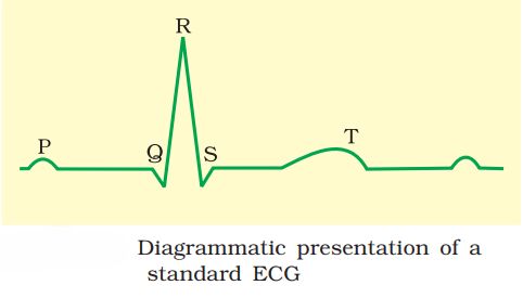
ECG is a graphical representation of the electrical activity of the heart during a cardiac cycle. To obtain a standard ECG (as shown in figure 18.3). a patient is connected to the machine with three electrical leads (one to each wrist and to the left ankle) that continuously monitor the heart activity. For a detailed evaluation of the heart’s function, multiple leads are attached to the chest region. Here, we will talk only about a standard ECG.
(1) Each peak in the; ECG is identified with a letter from P to T that corresponds to a specific electrical activity of the heart.
(2) The P-wave represents the electrical excitation (or depolarization) of the atria, which leads to the contraction of both the atria.
(3) The QRS complex represents the depolarization of the ventricles, which initiates the ventricular contraction. The contraction starts shortly after Q and marks the beginning of the systole.
(4) The T-wave represents the return of the ventricles from an excited to normal state (repolarisation). The end of the T-wave marks the end of the systole. Obviously, by counting the number of QRS complexes that occur in a given time period, one can determine the heartbeat rate of an individual. Since the ECGs obtained from different individuals have roughly the same shape for a given lead configuration, any deviation from this shape indicates a possible abnormality or disease. Hence it is of great clinical significance.
Additional Question and Answer
Question 1.
Explain the process of blood coagulation and the role of different components in it.
Answer:
Blood coagulation is the process by which blood forms a clot to prevent excessive loss after injury.
Steps and components involved:
- Injury to a blood vessel triggers platelets to release factors that initiate coagulation.
- Prothrombin, an inactive plasma protein, is converted into thrombin by the enzyme thrombokinase.
- Thrombin converts fibrinogen (plasma protein) into fibrin, forming a network of threads that trap blood cells.
- This fibrin mesh forms the clot, stopping blood loss.
Role of components:
- Platelets: Release clotting factors.
- Calcium ions (Ca²⁺): Essential for activation of clotting enzymes.
- Fibrinogen: Forms the fibrin mesh.
- Thrombin and thrombokinase: Catalyze the conversion of fibrinogen into fibrin.
Significance:
Prevents excessive blood loss and protects the body from infection.
Question 2.
What are the main functions of blood?
Answer:
Blood transports oxygen, carbon dioxide, nutrients, hormones, and waste products. It also helps in regulation of body temperature, immunity, and clotting.
Question 3.
Name the two major types of white blood cells (WBCs) and their functions.
Answer:
- Granulocytes: Neutrophils, Eosinophils, Basophils – involved in phagocytosis, infection resistance, and inflammatory response.
- Agranulocytes: Lymphocytes and Monocytes – responsible for immune responses and phagocytosis.
Question 4.
What is erythroblastosis fetalis? How can it be prevented?
Answer:
It is a condition where Rh-negative mother’s antibodies attack Rh-positive fetal RBCs, causing anemia or jaundice in the baby.
Prevention: Administer anti-Rh antibodies to the mother immediately after the first delivery.
Question 5.
Differentiate between systemic circulation and pulmonary circulation.
Answer:
- Pulmonary circulation: Right ventricle → lungs → left atrium. Carries deoxygenated blood to lungs for oxygenation.
- Systemic circulation: Left ventricle → body tissues → right atrium. Carries oxygenated blood to all body parts.
Question 6.
What are the layers of arteries and veins?
Answer:
- Tunica intima: Inner squamous endothelium.
- Tunica media: Middle layer of smooth muscle and elastic fibers.
- Tunica externa: Outer fibrous connective tissue.
(Veins have thinner tunica media than arteries.)
Question 7.
Explain the significance of coronary circulation.
Answer:
Coronary circulation supplies blood to the heart muscles (myocardium), providing oxygen and nutrients and removing wastes, essential for proper heart function.
Question 8.
What is the function of lymph?
Answer:
- Carries nutrients, hormones, and immune cells.
- Absorbs fats from the digestive system via lacteals.
- Drains tissue fluid back into major veins, maintaining fluid balance.
Question 9.
Define hypertension and mention one risk associated with it.
Answer:
Hypertension is persistently high blood pressure (≥140/90 mmHg).
Risk: Can lead to heart disease, stroke, or kidney damage.
Question 10.
What is the role of the nodal tissue in the heart?
Answer:
Nodal tissue (SAN, AVN, bundle of His, Purkinje fibers) generates and conducts action potentials, coordinating rhythmic contraction of the heart.
Question 11.
Why is the stroke volume important? How is cardiac output calculated?
Answer:
- Stroke volume: The volume of blood pumped by each ventricle per beat (~70 mL).
- Cardiac output: Stroke volume × Heart rate. Average ~5 L/min in a healthy adult.